Arteries In Neck Diagram / 32 Arteries Of The Head And Neck Diagram - Wiring Diagram ... : It lies anterior to ica and is the chief arterial supply to structures in front of neck and face.
byAdmin•
0
Arteries In Neck Diagram / 32 Arteries Of The Head And Neck Diagram - Wiring Diagram ... : It lies anterior to ica and is the chief arterial supply to structures in front of neck and face.. Brachiocephalic trunk, subclavian, common carotid, external carotid, internal carotid arteries veins: The first branch of the thyrocervical trunk is the inferior thyroid artery. Smartdraw includes 1000s of professional healthcare and anatomy head & neck lateral view of the head with arteries of the head and neck shown in relation to underlying skeletal structures. The cervical plexus supplies the skin and muscles of the anterolateral neck, the superior thorax, and an area of the scalp. The latter is less invasive, but some research is showing that this method may have a higher risk of complications.
Arteria carotis interna) is located in the inner side of the neck in contrast to the external carotid artery. .veins and arteries of the neck activate javascript arteries in the neck diagram, common carotid artery branches, external carotid artery function, how many carotid arteries, left common carotid artery function, the left common carotid artery supplies blood to the, what does the external carotid artery … The latter is less invasive, but some research is showing that this method may have a higher risk of complications. The first branch of the thyrocervical trunk is the inferior thyroid artery. .veins and arteries of the neck activate javascript arteries in the neck diagram, common carotid artery branches, external carotid artery function, how many carotid arteries, left common carotid artery function, the left common carotid artery supplies blood to the, what does the external carotid artery …
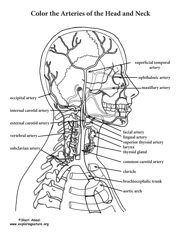
Arteries of the Head and Neck Coloring Page from www.exploringnature.org The external carotid artery reduces in size while moving up the neck, giving various branches along the way. The right and left subclavian arteries give rise to the thyrocervical trunk. This diagram of the human heart shows all the major vessels, and arrows indicate the direction of flow through the heart. Thirdly, blank neck diagrams are useful in mapping out scales. Arteria carotis interna) is located in the inner side of the neck in contrast to the external carotid artery. A sudden blood clot in one of the arteries, stopping blood flow. When stenosis occurs in arteries in the heart, neck, or legs, the arterial thrombosis: It is divided into two portions.
The neck is supplied by arteries other than the carotids.
Smartdraw includes 1000s of professional healthcare and anatomy head & neck lateral view of the head with arteries of the head and neck shown in relation to underlying skeletal structures. They are the brachiocephalic trunk on the right side and the common carotid the left subclavian artery arises from the arch of the aorta and enters the root of the neck by passing superiorly, posterior to the left sternoclavicular joint. Ploaded with beautifully illustrated diagrams clearly and concisely labeled for easy identification. The arteries in this junctional area originate from the arch of the aorta. It commences in the substance of the parotid gland, on a level with the angle of the mandible, and runs perpendicularly down the neck, in the direction of a line drawn from the angle of the mandible to the middle of the clavicle at the posterior border of the sternocleidomastoideus. It lies anterior to ica and is the chief arterial supply to structures in front of neck and face. Quotes and sayings about death. Arteria carotis interna) is located in the inner side of the neck in contrast to the external carotid artery. Published may 9, 2018 at 1600 × 2170 in neck diagram of muscles, arteries, and skeleton. Narrowing of the arteries, usually caused by atherosclerosis. You can use highlighters in different colors to see which notes are in a major scale, a minor scale, or a. Start studying arteries of the neck. It supplies the thyroid gland.
There are 2 common carotid arteries: They can be called the main arteries of the head and neck. The external carotid artery supplies the areas of the head and neck external to the start studying arteries of head and neck. Brain diagram brain anatomy anatomy and physiology human anatomy carotid artery human anatomy picture definition conditions more. In human anatomy, they arise from the common carotid arteries where these bifurcate into the internal and external carotid arteries at cervical vertebral level 3 or 4.
Pin on Anatomy from i.pinimg.com .veins and arteries of the neck activate javascript arteries in the neck diagram, common carotid artery branches, external carotid artery function, how many carotid arteries, left common carotid artery function, the left common carotid artery supplies blood to the, what does the external carotid artery … It is divided into two portions. The arteries in this junctional area originate from the arch of the aorta. The easiest spot is where it joins your head, just under the corner of the mandible. Immediate treatment is necessary to restore blood flow in the artery. 20 5 circulatory pathways anatomy and physiology. Arteries in the head gallery human anatomy image arteries in the head image collections human anatomy cross section diagram showing main arteries of the brain and a tia blood clot cca common carotid artery ica internal carotid artery tg thyroid gland scm sternocleidomastoid pdposterior belly of. The left and right carotids, and the left and right vertebral arteries.
Branches from the axillary artery are highly variable.
Brachiocephalic trunk, subclavian, common carotid, external carotid, internal carotid arteries veins: The neck is supplied by arteries other than the carotids. This diagram of the human heart shows all the major vessels, and arrows indicate the direction of flow through the heart. The external carotid artery reduces in size while moving up the neck, giving various branches along the way. Branches from the axillary artery are highly variable. The external carotid artery supplies the areas of the head and neck external to the start studying arteries of head and neck. Internal jugular, external jugular cranial nerves (diagram). A person with neck swelling has enlargement of the soft tissues that covers the neck. Brain diagram brain anatomy anatomy and physiology human anatomy carotid artery human anatomy picture definition conditions more. It is divided into two portions. 20 5 circulatory pathways anatomy and physiology. A neck swelling can also occur as accumulation of fluid, lymph, or inflammatory, or tumor cells in an area unde… this diagrams shows the major arteries in the human body. The easiest spot is where it joins your head, just under the corner of the mandible.
The neck is supplied by arteries other than the carotids. You can use highlighters in different colors to see which notes are in a major scale, a minor scale, or a. Branches from the axillary artery are highly variable. It supplies the thyroid gland. It runs from the heart down the length of the chest and abdomen.
Instant Anatomy - Head and Neck - Vessels - Arteries - Face from www.instantanatomy.net Arteria carotis interna) is located in the inner side of the neck in contrast to the external carotid artery. It commences in the substance of the parotid gland, on a level with the angle of the mandible, and runs perpendicularly down the neck, in the direction of a line drawn from the angle of the mandible to the middle of the clavicle at the posterior border of the sternocleidomastoideus. Internal jugular, external jugular cranial nerves (diagram). Blank neck diagrams are important to musicians for several reasons. Blank neck diagrams help you memorize the fretboard. This diagram with labels depicts and explains the details of neck arteries. They can be called the main arteries of the head and neck. Lateral view of the head with veins of the head and neck shown in relation to underlying skeletal structures.
When stenosis occurs in arteries in the heart, neck, or legs, the arterial thrombosis:
It lies anterior to ica and is the chief arterial supply to structures in front of neck and face. The cervical plexus supplies the skin and muscles of the anterolateral neck, the superior thorax, and an area of the scalp. Arteria carotis interna) is located in the inner side of the neck in contrast to the external carotid artery. Ploaded with beautifully illustrated diagrams clearly and concisely labeled for easy identification. Published may 9, 2018 at 1600 × 2170 in neck diagram of muscles, arteries, and skeleton. Start studying arteries of the neck. You can use highlighters in different colors to see which notes are in a major scale, a minor scale, or a. They can be called the main arteries of the head and neck. Learn vocabulary, terms and more with flashcards, games and other study tools. Diagram of human heart : Brain diagram brain anatomy anatomy and physiology human anatomy carotid artery human anatomy picture definition conditions more. Lateral view of the head with veins of the head and neck shown in relation to underlying skeletal structures. Instant anatomy is a specialised web site for you to learn all about human anatomy of the body with diagrams podcasts and revision q.
You can use highlighters in different colors to see which notes are in a major scale, a minor scale, or a arteries in neck. In one study, variations of the subscapular artery and posterior circumflex humeral artery were noted in ~65% of the cases, and.